Contact Info
Location
90 South Henry
Delaware, OH 43015

Confocal microscopy is revolutionizing many areas of study in biology. Through a process referred to as “optical sectioning,” confocal microscopes allow researchers to visualize samples without the need for destructive physical sectioning. Confocal microscopy also provides other advantages over transmitted and epifluorescence microscopy, including very high resolution 3D analysis of samples, and high-resolution time-lapse observations. To find out more about confocal microscopy, how it works, and how we’re using it at Ohio Wesleyan University, follow the links on the left.
A grant from the National Science Foundation made it possible for us to acquire an Olympus Fluoview 300 confocal microscope. With this addition to our already outstanding biological imaging facilities, we hope to provide all interested students with training on these cutting-edge instruments.
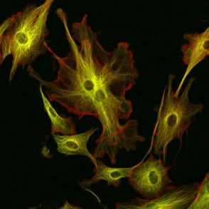
Bovine pulmonary arterial endothelial cells which have been dual labeled with a red stain for actin microfilaments (Rhodamine Phalloidoin) and a green fluorescent antibody against microtubules. These are the same cells as shown above, however the microtubules are found dispersed in the cytoplasm where they form an important part of the cytoskeleton.
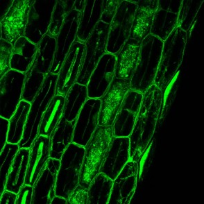
Basal region of an Arabidopsis root expressing GFP targeted to the vacuolar membrane. Obtained from ABRC stock center, stock number CS84727, as part of the Ehrhardt GFP collection.
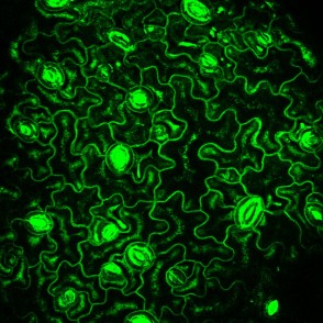
Arabidopsis leaf epidermis expressing GFP fused to an aquaporin that localizes to the plasma membrane (ABRC stock no. CS84758). A stoma is readily apparent at the lower right of the image. The “jigsaw” cells are known as pavement cells and make up the bulk of the cells of the epidermis.
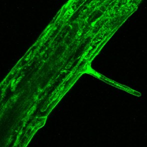
Arabidopsis root expressing GFP targeted to an unknown subcellular location (ABRC stock no. CS84729). Some appears to be localized to the endoplasmic reticulum. A root hair, formed by localized tip growth of an epidermal cell, is apparent in the middle of the image.
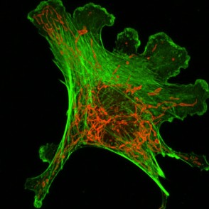
Bovine pulmonary arterial endothelial cells which have been dual labeled with a green stain for actin microfilaments (FITC-Phalloidoin) and a red stain for mitochondria. The stain for actin is actually derived from a toxic compound found in mushrooms. The stain for mitochondria is interesting because it only becomes fluorescent when the stain is activated by enzymes which reside in the mitochondria. For this reason, only the mitochondria appear red, not the other organelles.
